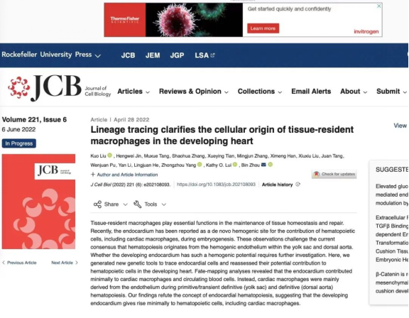
On April 28, 2022, theJournal of Cell Biologypublished online "Lineage tracing clarifies the cellular origin of tissue resident macrophages in the developing heart", the research results ofZhou Bin, Chief Professor of the School of Life Science, HIAS.

In this study, a variety of genetic lineage tracing systems were used to efficiently and specifically label endocardial cells, yolk sac (YS) endothelial cells, and aorta-gonads-mesonephros (AGM) endothelial cells in mice, revealing resident cardiac macrophages in the embryonic stage mainly originate from primitive hematopoiesis and transient definitive hematopoiesis of YS hemogenic endothelium and then definitive hematopoiesis of AGM hemogenic endothelium.

Note: Resident cardiac macrophages in the embryonic stage mainly originate from①primitive hematopoiesis and transient definitive hematopoiesis of YS hemogenic endothelium and②definitive hematopoiesis of AGM hemogenic endothelium rather than③cardiac endocardial cells.
As a "body engine", the heart is vital to maintaining human life activities. Cardiac dysfunction caused by a variety of cardiovascular diseases, such as myocardial infarction, myocardial inflammation, coronary heart disease, and hypertension, has severely threatened human health and become an important cause of human death. Resident cardiac macrophages are part and partial of the heart and play an important role in the metabolic homeostasis of the heart and cardiac regeneration. For instance, they can regulate cardiac coronary artery and lymphangiogenesis in the embryonic stage, maintain the normal function of the heart conduction system, and participate in the repair after cardiac injury. Therefore, elucidating the developmental origin of cardiac macrophages will provide an important research basis for cardiac development and clinical cardiac repair (Ginhoux and Guilliams, 2016; Grainger and Traver, 2019; Kim et al., 2020).
The developmental origin of resident cardiac macrophages has been scientifically controversial. Several studies have suggested that tissue-resident macrophages of mammalians in the embryonic stage originate from early hemogenic endothelium, mainly including hemogenic endothelial cells in YS and AGM (Ginhoux and Guilliams, 2016). There are mainly three rounds of hematopoiesis in the embryonic stage of mice. The first originates from the posterior plate mesoderm in YS blood island on Day 7.5 (E7.5) of embryonic development, producing early erythro-myeloid precursors (EMPs) that can differentiate into CD45+F4/80bright CD11 blow macrophages. The macrophages can migrate to the brain to form microglia, which is called primitive hematopoiesis. The second originates from the YS hemogenic endothelium on Day E8.0-E8.5 and can produce advanced EMPs. That is considered to be transient definitive hematopoiesis. Once the circulatory system is established around Day E8.5, EMPs colonize the fetal liver with the blood circulation and expand and differentiate into CD45+F4/80 low CD11bhigh macrophages/monocytes. The third originates from hematopoietic stem cells (HSCs) produced by the hemogenic endothelium in the AGM around Day E10.5, which is called definitive hematopoiesis. Those HSCs are transplanted into the fetal liver, continue to perform hematopoiesis around Day E10.5, and differentiate into various lineages of blood cells including CD45+F4/80lowCD11bhigh macrophages/monocytes (Ginhoux and Guilliams, 2016). The identification and discovery of new hemogenic endothelium has been a hot topic in developmental biology.
Recently, Haruko Nakano's group proposed a new concept that the endocardium also belongs to the hematopoietic endothelium. It can perform hematopoiesis in the early embryonic stage, and contribute to peripheral blood cells and differentiate into resident cardiac macrophages (Nakano et al., 2013; Shigeta et al., 2019). In their report, the endocardium was first labeled by using Nkx2.5-Cre(Nakano et al., 2013) to perform lineage tracing analysis. It was found that the endocardium transiently performs hematopoiesis around Day E9.5, and these endocardial-derived blood cells can enter the blood circulation. Later, the endocardial cells were labeled by using Nfatc1-Cre(Shigeta et al., 2019) to perform lineage tracing analysis, further indicating that endocardial cells can start to differentiate into cardiac macrophages around Day E9.5 and that this group of macrophages is very important for the development of heart valves. Removing them will cause abnormal valve development. It should be noted that in lineage tracing, the specificity of gene promoter expression will determine the specificity of reporter genes to target cell markers. For example, if the promoter of gene a is expressed in B cells in addition to A cells, the reporter gene will label both A and B cells in a-Cre;Rosa26-reporter, which will seriously interfere with the reliability of lineage tracing results (Tian et al., 2015). Therefore, strict specific marker detection is required for genetic tools before lineage tracing, which is an important prerequisite for the reliability of lineage tracing results. It has been found that Nkx2.5-Cre used to label the tracing of endocardial cells is controversial. Nkx2.5 is not a specific molecular marker for the endocardium, and Nkx2.5-Cre is expressed "ectopically" in the hemogenic endothelium of YS and AGM in addition to the endocardium (Zamir et al., 2017). Therefore, the scientific controversy of revealing whether Nfatc1-Cre specifically labels endocardial cells and elucidating the developmental origin of cardiac macrophages is urgent to be resolved.
In this study, the researchers of Zhou Bin's group first elaborated on the expression profile of Nfatc1. They built three kinds of Nfatc1, including Nfatc1-ires-Cre;R26-tdTomato,Nfatc1-2A-Dre;R26-RSR-tdTomato, and Nfatc1-2A-CreER;R26-tdTomato. By using the three kinds of Nfatc1 to perform systematic lineage tracing, they found that Nfatc1 is expressed in endocardial cells, epithelial cells in YS and AGM, and some peripheral blood immune cells, including macrophages, monocytes, neutrophils, T cells, and B cells. The above results reveal that Nfatc1 is not a specific molecular marker of the endocardium, and the peripheral blood macrophages and monocytes, as well as the hemogenic endothelium in YS and AGM it labels will greatly interfere with the endocardial lineage tracing results. Therefore, whether the endocardium belongs to the hemogenic endothelium and can perform hematopoiesis remains controversial. To resolve this controversy, they introduced two new lab mice in terms of the endocardium, including Mef2c-AHF-Cre; R26-tdTomato and Npr3-CreER; R26-tdTomato. Through systematic lineage tracing, it was confirmed that Mef2c-AHF-Cre could efficiently label atrial and outflow tract endothelial cells, while Npr3-CreER could label endocardium in an efficient and specific manner. Further experiments showed that none of these labeled endocardium has hematopoietic activity, nor do they differentiate into resident cardiac macrophages or label peripheral blood cells. On the other hand, researchers also ruled out the possibility that the epicardium has the potential to perform hematopoiesis by specifically labeling epicardial cells with Wt1-CreER; R26-tdTomato. Therefore, in order to re-elucidate the developmental origin of resident cardiac macrophages, they constructed Cdh5-2A-CreER and validated its ability to efficiently and specifically label hematopoietic endothelium. Inducing E7.5Cdh5-2A-CreER;R26-tdTomato by 4OHT can efficiently trace brain microglia, suggesting that the primitive hematopoiesis and transient definitive hematopoiesis of the hematopoietic endothelium of YS are specifically and efficiently captured. By contrast, inducing E10.5 Cdh5-2A-CreER;R26-tdTomato by 4OHT cannot trace brain microglia, suggesting that the definitive hematopoiesis of hematopoietic endothelium of AGM is specifically and efficiently captured without interference from YS hematopoietic endothelium. Then, the embryonic hearts of E16.5Cdh5-2A-CreER;R26-tdTomato were collected for analysis to reveal that resident cardiac macrophages during the embryonic stage originate primarily from the primitive hematopoiesis and transient definitive hematopoiesis of the hematopoietic endothelium of YS (approximately 90%) and then from the definitive hematopoiesis of hematopoietic endothelium of AGM (approximately 15%). This study illustrates that the developmental origin of resident cardiac macrophages will provide an important research basis for cardiac development and clinical immunotherapy of cardiac diseases.
Liu Kuo, postdoctoral fellow of the research group of Zhou Bin, School of Life Science, HIAS, and Jin Hengwei, postdoctoral fellow of the Center for Excellence in Molecular Cell Science, CAS, are the co-first authors of the paper. Professor Zhou Bin from HIAS is the corresponding author of the paper.The study is strongly supported by the laboratory animal platform and cell analysis technology platform of CEMCS and the National Facility for Protein Science Shanghai(NFPSS). The work is funded by the Chinese Academy of Sciences, the National Natural Science Foundation of China, the Ministry of Science and Technology of the People's Republic of China, and the Science and Technology Commission of Shanghai Municipality.
Source | School of Life Science
Typesetter | Zhou Liyuan
Published in Zhejiang
Read More

Scan with WeChat
Follow us